
Introduction To C-Arm glTF
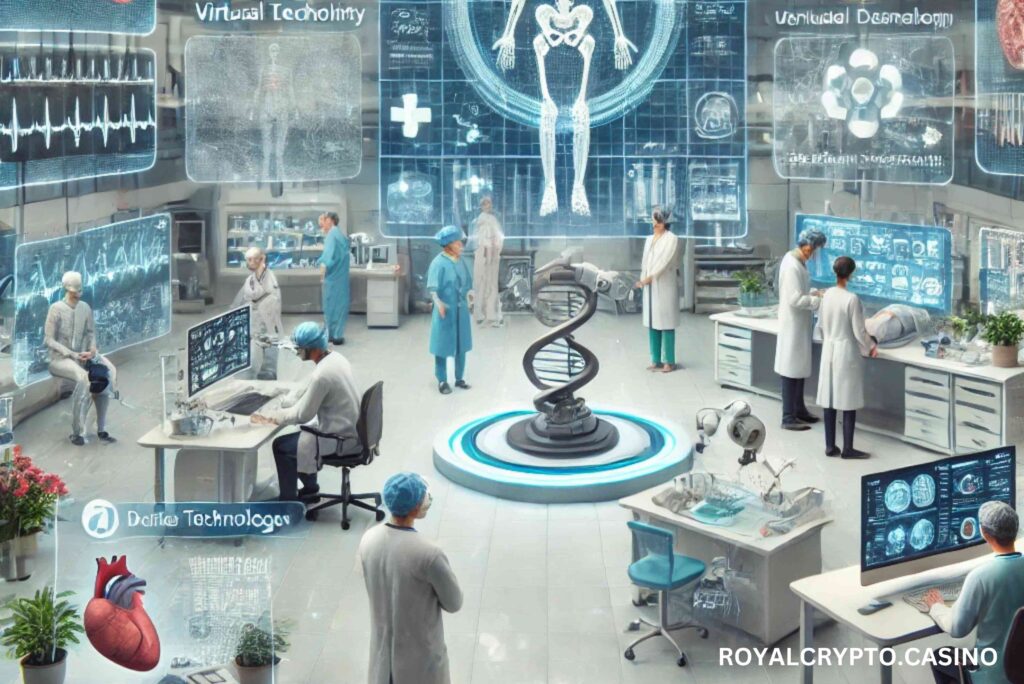
So, what exactly is glTF? Short for Graphics Language Transmission Format, glTF is like the JPEG of 3D models. c-arm gltf It’s a lightweight file format that makes sharing and visualizing 3D content seamless. Developed by the Khronos Group, glTF is widely used in industries like gaming and virtual reality. Now, it’s making its way into healthcare.
The beauty of glTF lies in its efficiency. It compresses complex 3D models without sacrificing quality, ensuring smooth, high-quality visuals. For medical imaging, this means doctors can explore highly detailed anatomical structures with ease.
The Fusion of C-Arm and glTF
Imagine combining the real-time imaging capabilities of a C-Arm with the 3D visualization power of glTF. This fusion creates a cutting-edge tool that offers unparalleled insights into the human body. By integrating glTF visualization into C-Arms, doctors can view detailed 3D models alongside live X-ray images, enhancing their ability to diagnose and treat conditions.
This combination is like upgrading from a paper map to a GPS system. It provides a clearer, more interactive view, making complex procedures more manageable.
Benefits of C-Arm glTF Visualization
- Enhanced Accuracy
With glTF’s high-quality 3D models, surgeons can pinpoint anatomical structures with incredible precision. This reduces the margin for error and leads to better outcomes.
- Real-Time Interaction
Doctors can manipulate 3D models in real time, zooming in on specific areas or rotating the view for a better angle. This level of interaction is a game-changer.
- Improved Communication
Complex medical information becomes easier to understand when visualized in 3D. Patients and medical teams can better grasp what’s happening, leading to more informed decisions.
- Cost-Effective Solutions
While advanced imaging technologies can be expensive, integrating glTF into existing C-Arms is a cost-efficient way to enhance their capabilities.
Applications in Modern Medicine
- Orthopedic Surgery
From fixing fractures to placing implants, C-Arm glTF visualization provides a clear view of bones and joints, making orthopedic procedures more precise.
- Cardiovascular Interventions
In heart surgeries and catheter placements, the technology helps visualize arteries and veins, ensuring accurate treatment.
- Neurological Procedures
Navigating the brain’s intricate structures becomes easier with detailed 3D models, reducing risks during delicate surgeries.
How It Enhances Surgical Precision
Precision is everything in surgery, and C-Arm glTF visualization delivers it in spades. By overlaying 3D models on live imaging, surgeons get a comprehensive view of the area they’re working on. This reduces guesswork and ensures that every move is deliberate and calculated.
Think of it as having a GPS guide you through a dense forest. You’re not just guessing your way forward; you have a clear path laid out in front of you.
Improving Patient Outcomes
When medical teams can see better, they can do better. C-Arm glTF visualization minimizes complications, shortens surgery times, and speeds up recovery. For patients, this means less pain, fewer risks, and a quicker return to normal life.
Challenges and Limitations
No technology is perfect, and C-Arm glTF visualization is no exception. Some challenges include:
- Integration Costs: Upgrading existing systems can be expensive.
- Learning Curve: Medical professionals need training to use the technology effectively.
- Technical Limitations: Not all hospitals have the infrastructure to support advanced visualization tools.
The Future of Medical Imaging
The journey of medical imaging is just beginning. As C-Arm glTF visualization becomes more widespread, we can expect even greater advancements. Think AI-powered imaging, augmented reality overlays, and even more accurate diagnostics. There are countless opportunities; the future seems bright.
FAQs About C-Arm glTF Visualization
- What is C-Arm glTF visualization?
It’s the integration of glTF 3D visualization technology with C-Arm imaging devices, offering real-time, high-quality insights for medical procedures.
- How does it benefit surgeons?
It enhances precision by providing detailed, interactive 3D models alongside live imaging, reducing errors and improving outcomes.
- Is it expensive to implement?
While there are upfront costs for integration and training, the long-term benefits often outweigh the expenses.
- Can it be used in all types of surgeries?
It’s particularly beneficial for orthopedic, cardiovascular, and neurological procedures but has potential applications across various fields.
- What does the future hold for this technology?
The future includes advancements like AI integration, augmented reality features, and broader accessibility, making medical imaging even more revolutionary.
